PL Projection: 3D Movie
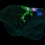
The marmoset brain with tracer injection to A46 was processed by serial two-photon tomography. This image stack shows serial sections of a marmoset brain that was processed in coronal direction. The red color represents the tissue autofluorescence and the green color represents the tracer signals. (by Yamamori Lab.) Video showcasing 3D volumetric of Marmoset brain with several tracers (AAV-GFP, AAV-Tom, Fast Blue) progression from injection site to terminal projection sites