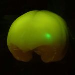
Marmoset Tracer Projects
Marmoset brain mapping based on tracer injection
Brain/MINDS is making a connection map of the marmoset cortex using anterograde tracer injection with virus vectors. In the first phase of the project (2014-2018), three groups, led by Tetsuo Yamamori, Noritaka Ichinohe, and Partha Mitra, were involved. The Yamamori team used serial two-photon tomography (STPT) for imaging the projections while Ichinohe and Mitra groups both used a digital slide scanner. For accurate 3D registration, all of the tracer-injected brains were imaged with an MRI-scanner before sectioning. The tracer signals were mapped to a reference 3D space, which will be used for integration of cross-modal experiments, including MRI, DTI and histological staining, in addition to the tracer data. In the second phase of the project (2019-2023) the tracer injection work has been consolidated and is continued on by Yamamori Team.
Features
-
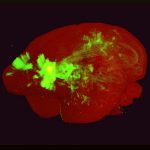
PFC Projection: 3D Movie
An example movie to demonstrate the axonal projections from the prefro...
-

TL Projection: 3D Movie
A sample of the 3D reconstructed marmoset atlas from tracer images.
-
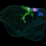
PL Projection: 3D Movie
The marmoset brain with tracer injection to A46 was processed by seria...
-
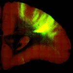
PFC Projection: Section Images
Image stacks for coronal sections of PFC projections
-
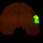
TL Projection: Section Images
Axonal projection maps in the whole marmoset brain.
-
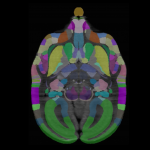
PL Projection: Section Images
Fully interactive portal site with user friendly options. Ability to n...
