Structural and Functional Map
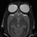
MRI is able to collect image data reflecting structural and functional information of the brain in vivo, non-invasively. Proton density and the amount of water are reflected in the relaxation time of each proton nucleus. Brain structural images are derived from the frequency domain representation. By adjusting the data collection parameters, we can collect different types of contrast images such as T1WI and T2WI. Functional MRI (fMRI) can capture neuronal activity through Blood oxygenation level dependent (BOLD) signal that reflects the degree of deoxidization of hemoglobin in the blood, correlated with neuronal activity.
Features
-

Marmoset Tracer Experiment
TissueCyte tracer experiment data.